General Description
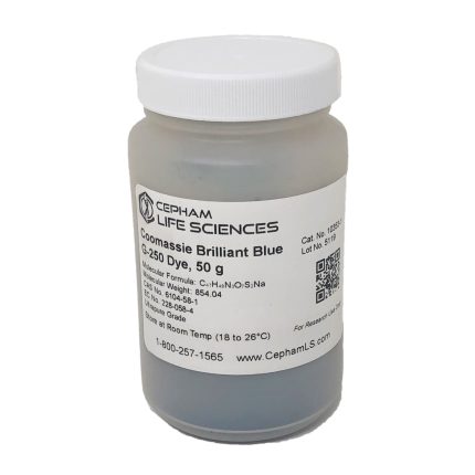
Coomassie brilliant blue staining is one of the most common methods of detecting proteins in polyacrylamide gels (PAGE).
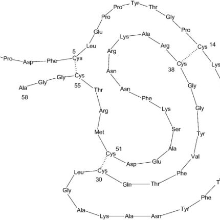
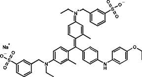
Two chemical forms of a disulfonated triphenylmethane compound commonly serving as the basis of stains used for detection of proteins in gel electrophoresis and Bradford-type assay reagents for protein quantitation methods are Coomassie R-250 and G-250 dyes. The Coomassie R-250 (develops red-tinted hue) lacks two methyl groups that are present in the G-250 (develops green-tinted hue) or Colloidal coomassie dye form. Coomassie gel stains and protein assay reagents are usually formulated as highly acidic solutions in methanol. Under such acidic conditions, the mechanism of dye binding to proteins is primarily facilitated by basic amino acids (primarily arginine, lysine and histidine), while the number of protein bound Coomassie dye ligands is approximately proportional to the number of positive charges found on the protein. Protein-binding changes the dye from reddish-brown to bright blue color with an absorption maximum at 595 nm.
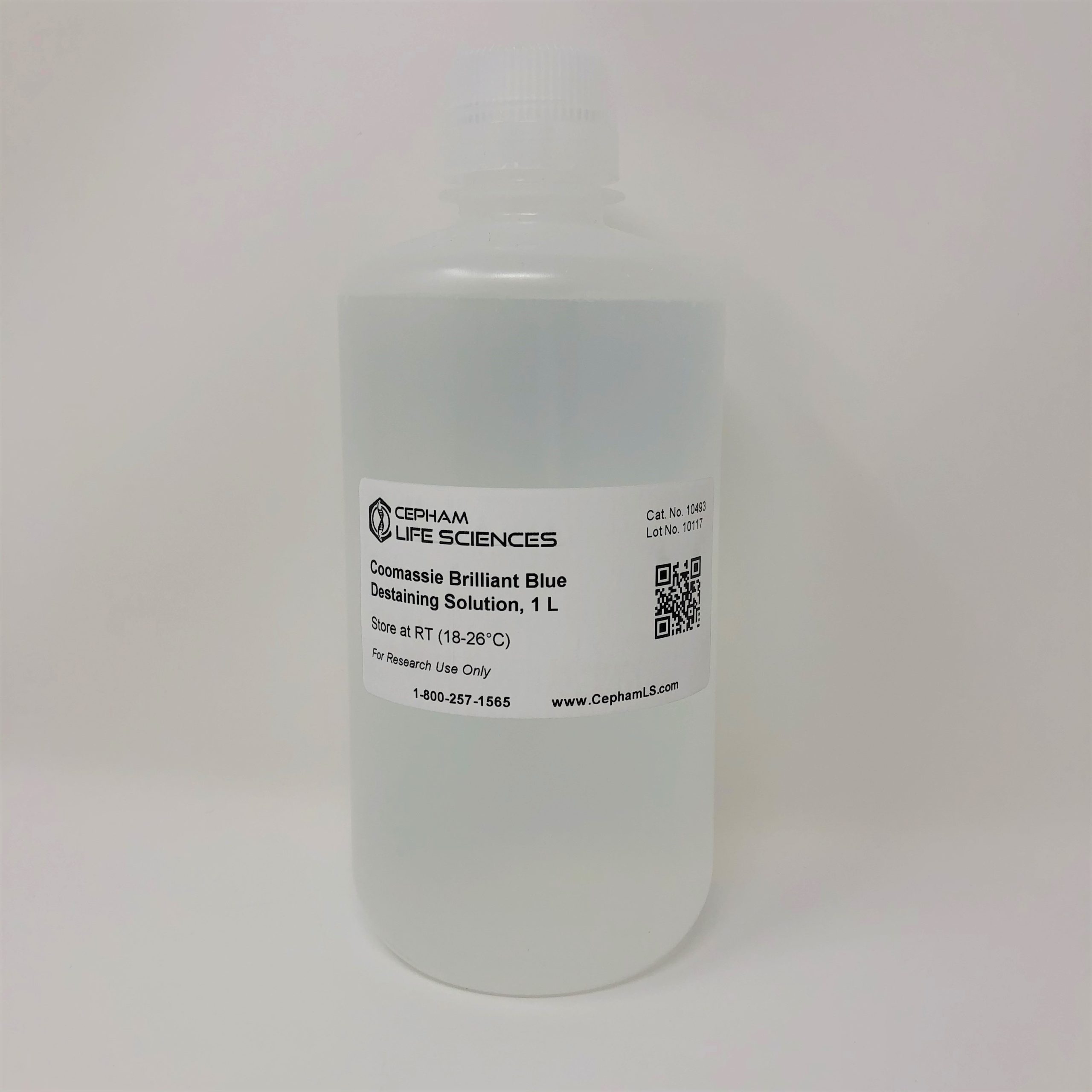
The Coomassie Brilliant Blue Destaining Solution is useful to remove excess stain, which allows for better visualization of proteins as blue bands on a clear background.




![IEF Anode Buffer [50X]](https://www.cephamls.com/wp-content/uploads/2019/02/10496-3-430x334.jpg)
