


Exosome Isolation Reagent Comparison Guide

Selecting an exosome isolation reagent or extracellular vesicle (EV) separation method should begin with the downstream application, not simply with the goal of maximizing particle recovery. A method that provides high particle recovery from conditioned cell culture medium, for example, may be less suitable for proteomic analysis if it also recovers abundant soluble proteins and other non-vesicular components.
Conversely, a method producing a lower apparent yield may be preferable when sample purity, RNA analysis, protein characterization, or functional studies are the primary objectives.
An important consideration is terminology. The International Society for Extracellular Vesicles (ISEV) recommends using extracellular vesicle (EV) as the general term for lipid-bilayer-delimited particles released from cells. The term exosome more specifically refers to EVs of endosomal origin and is most appropriate when that origin can be demonstrated. However, “exosome isolation” remains widely used in laboratory and commercial terminology to describe workflows intended to isolate or enrich small EV populations.
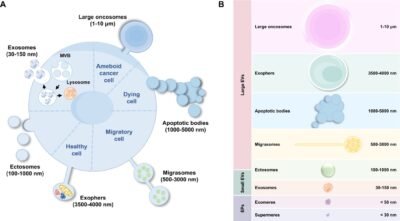
Extracellular vesicle diagram showing exosomes and other EV types
[Image: Sai Chitti, Wikimedia Commons, CC BY 4.0.]
What to Compare in Exosome Isolation Reagents
No single EV isolation method is optimal for every experiment. Before selecting a kit or reagent, compare the expected performance across several practical criteria: starting sample type and volume, recovery yield, preparation purity, processing time, sample throughput, equipment requirements, and compatibility with the planned downstream assay.
Sample type can substantially affect method performance. Conditioned cell culture medium is generally less complex than plasma, serum, urine, cerebrospinal fluid, or other biological fluids. Protein-rich samples may contain lipoproteins, protein aggregates, and other non-vesicular extracellular particles that can co-isolate with EVs. A method that performs well with conditioned medium should therefore not automatically be assumed to perform equally well with a more complex biological sample.
Yield should also be interpreted carefully. Particle number, total protein, total RNA, and EV-associated marker abundance measure different characteristics of a preparation. A high total protein concentration, for example, does not necessarily indicate high EV recovery because soluble proteins and other contaminants may contribute to the measurement.
For this reason, EV preparations are best evaluated using complementary characterization methods appropriate to the research question and downstream application.
Exosome Isolation Reagent Comparison by Method Type
Polymer Precipitation Reagents
Polymer-based precipitation reagents are widely used because they provide a relatively simple workflow and generally do not require ultracentrifugation. These reagents alter the solubility of EV-containing material, allowing vesicles and associated material to be recovered using comparatively low-speed centrifugation.
Precipitation-based methods can be useful when processing multiple samples, working with limited sample volumes, maximizing recovery, or when access to ultracentrifugation equipment is limited.
The principal limitation is co-precipitation. Soluble proteins, protein complexes, lipoproteins, nucleic acids, and other components may be recovered along with EVs.
This becomes particularly important in proteomics and biomarker research, where abundant contaminating proteins can interfere with the detection of lower-abundance EV-associated proteins.
For Western blotting, ELISA, or preliminary RNA studies, precipitation methods may nevertheless provide a practical workflow when appropriate controls and, where necessary, additional cleanup steps are incorporated.
Affinity Capture Reagents
Affinity-based reagents use specific molecular interactions to enrich extracellular vesicles or selected EV populations. Depending on the reagent and isolation chemistry, these interactions may involve membrane-associated molecules or other characteristics of the vesicle surface.
The primary advantage of affinity-based isolation is selectivity.
The trade-off is that affinity isolation may enrich only the EV population carrying the targeted characteristic. The resulting preparation therefore may not represent the complete EV population present in the original sample.
This can be useful when the objective is targeted enrichment, but it may be less appropriate when broad EV profiling is required.
Researchers should also consider whether the binding and elution conditions are compatible with downstream applications such as RNA extraction, Western blotting, mass spectrometry, immunoassays, or functional studies.
Size-Exclusion Cleanup and Reagent-Supported Workflows
Size-exclusion chromatography (SEC) separates particles according to hydrodynamic size and can help reduce contamination from smaller soluble proteins.
SEC can be useful when cleaner preparations are required for sensitive downstream analysis. It may also be combined with a concentration step to achieve an appropriate balance between recovery and purity.
However, size-based separation cannot completely distinguish EVs from other particles of similar size. EVs and non-vesicular extracellular particles can have overlapping physical characteristics, particularly in complex biological samples.
Another practical consideration is sample dilution. EV-containing fractions may require concentration before protein analysis, RNA isolation, or functional assays.
For some applications, a combined workflow—such as an initial concentration step followed by size-exclusion cleanup—may provide a more suitable preparation than either approach alone.
Immunocapture Reagents
Immunocapture methods use antibodies directed against selected EV-associated antigens to enrich a defined vesicle population.
This approach can be particularly useful when the experimental objective is the detection or analysis of EVs displaying a particular surface marker rather than recovery of the total EV population.
Important variables include antibody specificity, epitope accessibility, bead or solid-phase chemistry, nonspecific binding, elution conditions, and the use of appropriate controls.
Because immunocapture selects a targeted population, the isolated fraction should not automatically be considered representative of all EVs present in the original sample.
Match the Reagent to the Downstream Assay
RNA Analysis
For RNA analysis, researchers should consider both RNA recovery and the presence of compounds that may interfere with reverse transcription or PCR.
Higher total RNA yield does not necessarily indicate greater EV-associated RNA recovery. Non-vesicular RNA and other co-isolated components can contribute to the measured RNA concentration.
Where appropriate, spike-in controls, replicate processing, and consistent sample handling can help evaluate extraction efficiency and reproducibility.
Western Blotting and Immunoassays
Western blotting is frequently used as one component of EV characterization.
Proteins commonly evaluated in EV studies include CD9, CD63, CD81, TSG101, and ALIX. However, the presence of an individual marker alone does not establish the identity or purity of an EV preparation.
Marker selection should be appropriate for the EV source and experimental design, and evaluation of potential non-EV contaminants should also be considered. Using multiple complementary approaches provides a more informative assessment than relying on a single marker.
Proteomic Analysis
For proteomic analysis, reducing contaminant burden may be more important than maximizing total particle recovery.
Polymer carryover, abundant serum proteins, and other co-isolated material can interfere with the detection of lower-abundance proteins. Appropriate cleanup procedures, process blanks, and consistent sample normalization can therefore be particularly important.
Protein extraction, purification, quantitation, electrophoresis, and Western blotting may all become part of the downstream workflow when EV preparations are being investigated at the protein level.
Functional Studies
When isolated EV preparations are applied to recipient cells, residual isolation reagents or co-isolated components may potentially affect cell viability, proliferation, apoptosis, migration, cellular uptake, signaling, or other biological responses.
Reagent-only or process controls should therefore be considered when evaluating these outcomes.
If an observed biological effect is also detected in an appropriately processed reagent control, the isolation procedure itself may be contributing to the measured response.
Build a Reproducible Evaluation Plan
Rather than selecting an isolation method solely on the basis of reported recovery, researchers can perform a small qualification study using the sample type and downstream application relevant to their experiment.
Where practical, identical aliquots can be processed using two or more candidate methods while keeping important pre-analytical variables consistent. These variables may include sample collection conditions, storage temperature and duration, freeze-thaw history, clarification procedures, starting sample volume, reagent-to-sample ratio, incubation conditions, centrifugation parameters, final resuspension volume, and downstream analytical method.
For an RNA-focused workflow, useful comparison criteria might include particle recovery, RNA yield, PCR inhibition, and target reproducibility.
For a protein-focused workflow, researchers may instead evaluate particle-to-protein relationships, EV-associated marker enrichment, background protein levels, and Western blot performance.
Recording reagent lot numbers, reagent-to-sample ratios, incubation times, centrifugation conditions, and final sample volumes can also help improve reproducibility between experiments.
Choosing the Appropriate EV Isolation Approach
There is no universally optimal exosome isolation reagent or EV separation method. The appropriate choice depends on the sample matrix, experimental objective, required purity, desired recovery, available equipment, sample throughput, and downstream assay.
Precipitation-based reagents can provide convenient and scalable recovery. Affinity and immunocapture approaches can provide greater selectivity for particular EV populations. Size-exclusion methods can help reduce some soluble contaminants, while combined approaches may provide a useful balance between recovery and sample purity.
Most importantly, EV isolation should be considered as part of the complete experimental workflow rather than as an isolated laboratory step. Evaluating the isolation method in relation to the intended downstream analysis can help researchers obtain more interpretable and reproducible results.
Cepham Life Sciences supports life science researchers with products for protein extraction and purification, protein quantitation, electrophoresis, Western blotting, ELISA, antibodies, biological buffers and reagents, and molecular biology workflows that can support downstream analysis of EV-containing research samples.
For Research Use Only.
References
- Welsh JA, Goberdhan DCI, O’Driscoll L, et al. Minimal information for studies of extracellular vesicles (MISEV2023): From basic to advanced approaches. Journal of Extracellular Vesicles. 2024;13(2):e12404. doi:10.1002/jev2.12404.
- Théry C, Witwer KW, Aikawa E, et al. Minimal information for studies of extracellular vesicles 2018 (MISEV2018): a position statement of the International Society for Extracellular Vesicles and update of the MISEV2014 guidelines. Journal of Extracellular Vesicles. 2018;7(1):1535750. doi:10.1080/20013078.2018.1535750.