


Cell Counter
Original price was: $5,800.00.$3,480.00Current price is: $3,480.00.
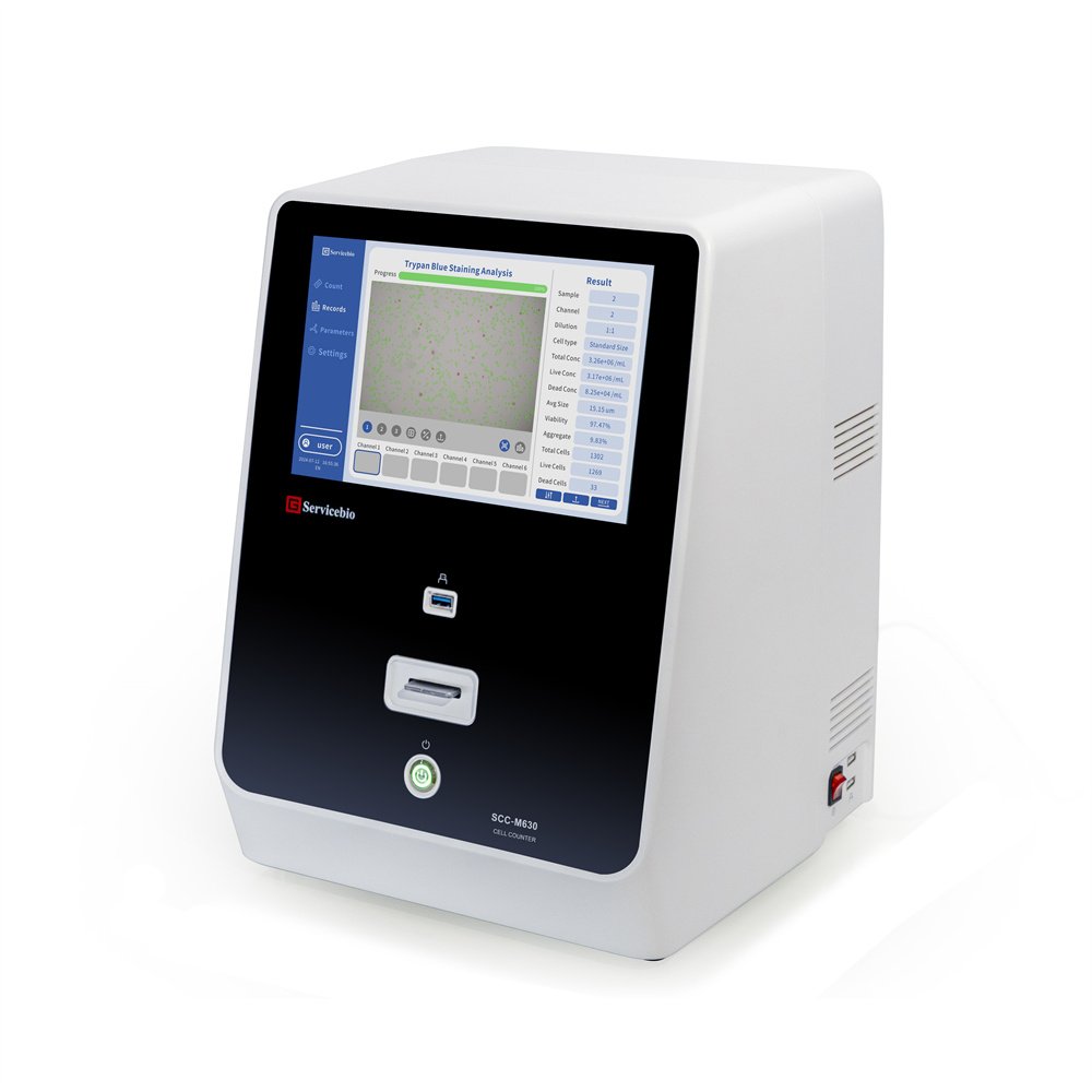
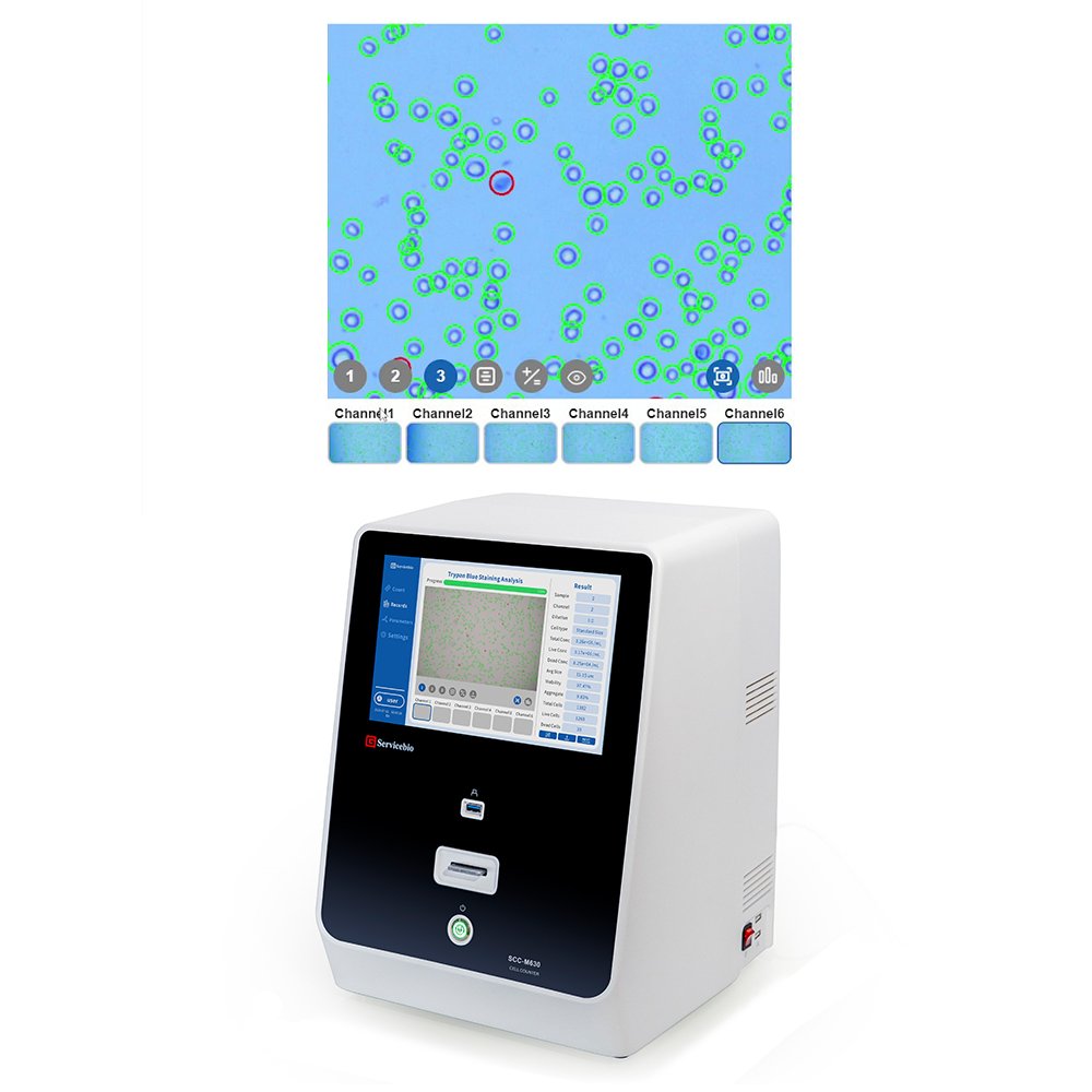
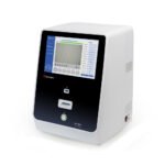
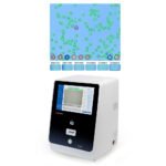
The cell counter is a precise and rapid cell analysis system that combines image recognition technology and optical imaging technology. It can obtain data such as cell number, concentration, and viability with one click, and display the morphology of cells.
The cell counter is a precise and rapid cell analysis system that combines image recognition technology and optical imaging technology. It can here obtain data such as cell number, concentration, and viability with one click, and display the morphology of cells.
Product Features
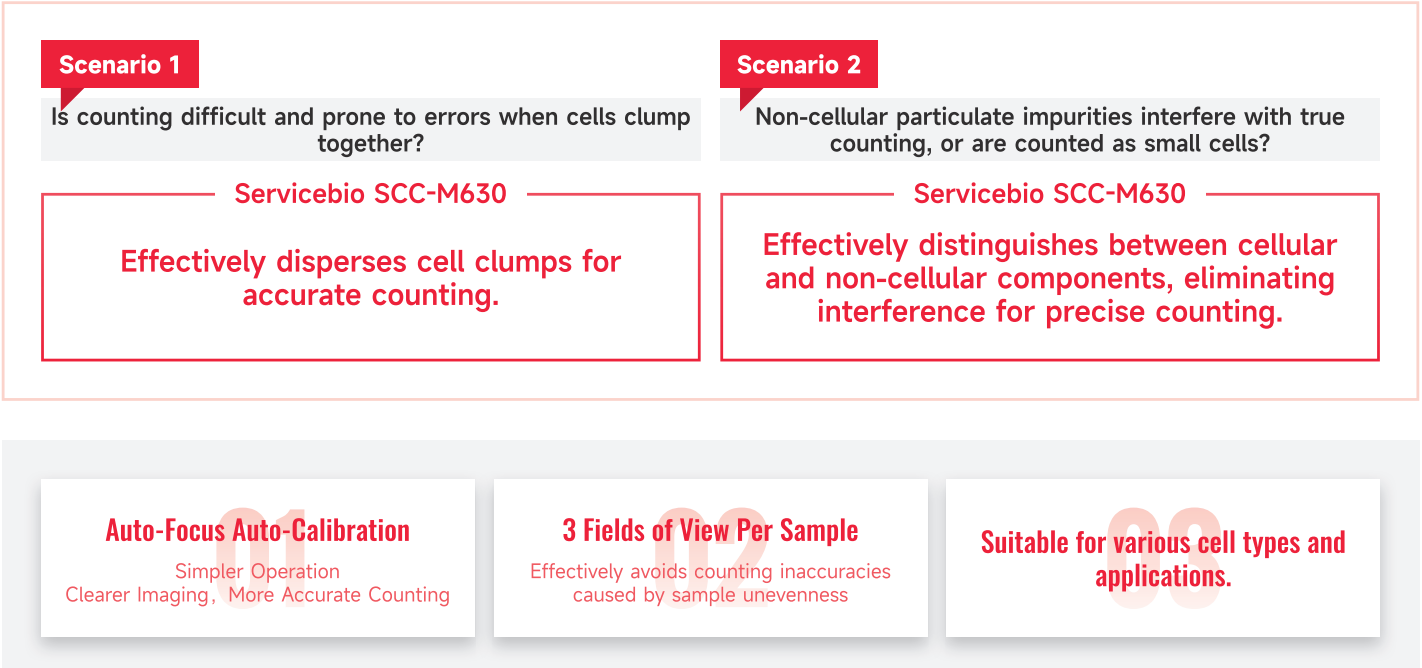
1. It can effectively break down clumps of cells for accurate counting.
2. It can effectively identify cellular and non-cellular components, eliminate interference, and ensure accurate counting.
3. It features automatic focusing and automatic calibration.
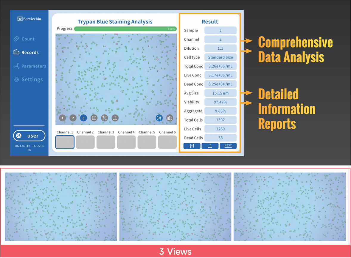
4. It provides 3 fields of view for each sample.
5. It is suitable for various cell types and application fields.
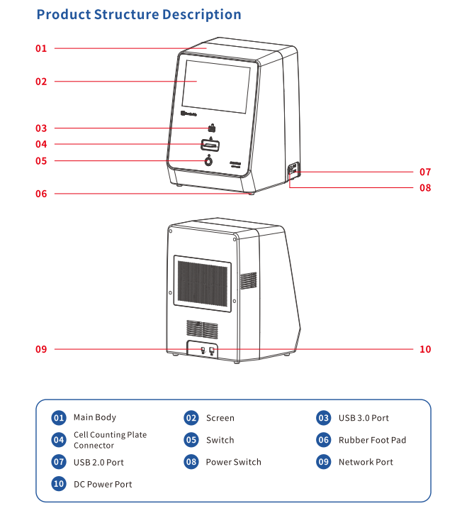
6. With 6-well high-throughput design, it offers fully automatic and precise counting.
7. It provides comprehensive data analysis and detailed information reports.
Product Parameters:
| Product name | Cell Counter |
| Cat.No. | SCC-M630 |
| Dimensions | 287x283x388 mm |
| NW | 9.6kg |
| Power | 70W |
| Electrical parameters | 12V |
| Operating temperature | 10-40°C |
| Ambient humidity | <80% |
| Consumable type | Disposable counting chamber |
| Single detection throughput | 6 times |
| Consumable throughput | 6-channel |
| Counting mode | Bright field/Trypan blue staining |
| Focusing method | Auto-focus |
| Sample introduction method | Automatic sample feeding |
| Pixel of material machine for pasting group | 6.3 million CMOS |
| Bright field light source | LED light |
| Optical magnification | 3X magnification |
| Field of view image | Single channel with 3 views of photography |
| Display module resolution | LCD 10.1″ multi-touch 1280X800 |
| Image resolution | 3072×2048 |
| Image format | JPG |
| Result output format | JPG/PDF/CSV |
| Storage space | 256G |
| Screen size | 10,1″ |
| Storage record | 1000 pcs |
| Sample type | Suspension-cultured cells/Adherent-cultured cells/Mammalian cells Human cells/PBMC and other cells |
| Cell concentration | 5×10⁴-1.5×10⁷ cells/mL |
| Optimal concentration range | 5×10⁵-1×10⁷ cells/mL |
| Cell diameter | 5-60μm |
| Counting time | Single channel < 205 |
| Sample volume | 10 μL |
Citation(s):
Tian, L., et al. PTTG1 promotes M2 macrophage polarization via the cGMP-PKG signaling pathway and facilitates EMT progression in human epithelial ovarian cancer cells. Discov Oncol 16, 730 (2025). PMID 40353994 IF 2.8
[vc_row][vc_column][vcex_button url=”`{`acf field=“safety“`}`” size=”large” text_source=”custom_text” aria_label=”Safety Datasheet” target=”blank” icon_left=”ticon ticon-file-pdf-o” icon_left_padding=”10px”]Safety Datasheet[/vcex_button][/vc_column][/vc_row]
Related products
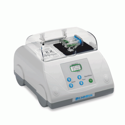
BEADBUG 3 MICROTUBE HOMOGENIZER, 115V
Original price was: $1,022.95.$869.51Current price is: $869.51.
Carousel Stand for Nichipet Air, Premium LT, Premium and LE Pipettes
Original price was: $115.00.$97.75Current price is: $97.75.