


Mammalian Cell Culture Contamination Control

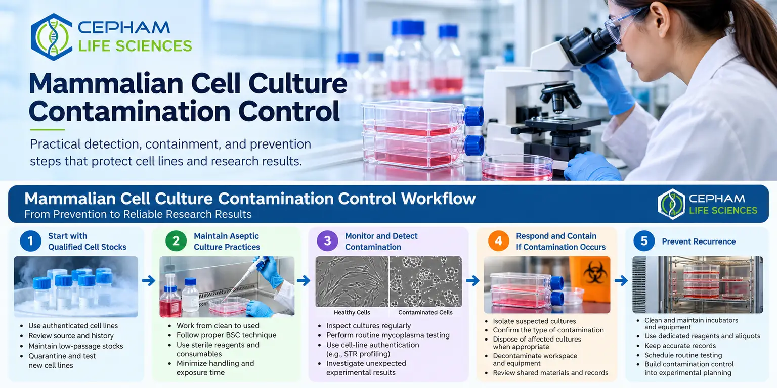
A cell line that grows slightly slower, detaches more readily, or produces an unexpected assay result may be contaminated long before visible changes appear. Mammalian cell culture contamination can compromise protein expression, transfection efficiency, signaling studies, cell viability measurements, and downstream molecular analysis. The most costly events are often not obvious bacterial outbreaks, but low-level mycoplasma contamination or cell-line cross-contamination that persists across multiple experiments.
For research laboratories, contamination control is not a single cleanroom practice or a reagent choice. It is a workflow that begins with qualified cell stocks, continues through daily aseptic handling, and includes routine surveillance and documented corrective actions.
What Mammalian Cell Culture Contamination Looks Like
Contamination is commonly divided into microbial contamination and cross-contamination with another cell line. The detection method, response, and experimental risk differ substantially between these categories.
Bacteria and fungi may cause rapidly developing turbidity, color changes in pH indicator-containing medium, floating particles, or a sharp decline in cell health. Yeast can appear as small, budding particles and may create visible cloudiness. Mold is often more apparent, with filamentous structures that can be seen under low magnification. These events are disruptive, but they are usually recognized quickly.
Mycoplasma presents a different problem. These organisms lack a cell wall, are small enough to evade standard light-microscopy detection, and may not cause visible turbidity. Contaminated cultures can remain apparently usable while showing altered metabolism, gene expression, growth rate, morphology, cytokine production, or response to treatment. Antibiotics may suppress visible evidence without eliminating the organism, which is one reason routine testing is more reliable than relying on appearance alone.
Cross-contamination occurs when cells from one culture are introduced into another. Fast-growing lines can overtake slower cultures, while closely related lines may be difficult to distinguish by morphology. This problem can invalidate months of work even when cultures are free of microorganisms. Cell-line authentication and careful culture segregation are therefore integral parts of contamination control.
Common Sources of Contamination in Cell Culture
Most contamination events arise from a limited number of recurring sources. Identifying the likely entry point helps laboratories correct the process rather than repeatedly discarding cultures without changing the underlying cause.
Personnel are a frequent source. Incomplete disinfection of gloves, hands positioned over open vessels, rapid movements that disrupt cabinet airflow, and handling nonsterile items near open cultures can all introduce contaminants. A biological safety cabinet supports aseptic technique, but it cannot compensate for poor cabinet practices or an obstructed airflow path.
Reagents and consumables are another consideration. Contamination may be introduced through improperly handled media bottles, shared aliquots, nonsterile water, compromised supplements, or pipettes that contact nonsterile surfaces. Serum, growth factors, and other biological components should be evaluated within the laboratory’s incoming-material and lot-management process. Repeatedly warming a shared stock can also increase handling events and create opportunities for contamination.
Incubators deserve particular attention because high humidity, warmth, and frequent access favor microbial growth. Water pans, shelves, door gaskets, and spilled medium can become persistent sources. Contamination can also spread through shared centrifuges, aspirators, microscope stages, and cryogenic storage practices when cleaning and sample organization are inconsistent.
Early Detection Protects Experimental Data
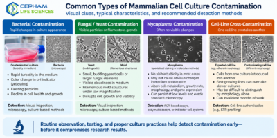
Visual inspection remains useful, but it should be performed systematically. Before feeding or passaging a culture, assess medium color, clarity, particulate matter, confluence, morphology, attachment, and the presence of floating cells. Compare the culture with documented expectations for that cell line rather than relying on a general impression that it “looks fine.”
Routine mycoplasma testing should be scheduled based on culture volume, project risk, and laboratory traffic. PCR-based assays can provide sensitive detection of mycoplasma nucleic acid, while other approaches detect enzymatic activity or use indicator-cell systems. The appropriate method depends on the laboratory workflow, turnaround requirement, and validation expectations.
Testing is particularly prudent when a new cell line enters the laboratory, after receipt from another group, following thaw from an older stock, before critical experiments, and at regular intervals during extended culture.
Cell-line authentication should also be built into the workflow. Short tandem repeat profiling is commonly used for human cell lines and can confirm identity or identify mismatch concerns. Authentication does not replace mycoplasma testing, and mycoplasma testing does not establish cell identity. Both controls address different risks.
When unexpected assay variability appears, contamination should be considered alongside other technical causes. A shift in Western blot signal, abnormal ELISA background, inconsistent transfection performance, or unexplained viability loss may originate in the culture system rather than the assay reagents or instrument settings.
Responding to a Suspected Contamination Event
A suspected culture should be isolated immediately. Do not continue routine use while waiting for confirmatory testing, and do not open the vessel alongside clean cultures. Label the material clearly, record the date and observed findings, and review which media, reagents, equipment, and cultures were shared.
For confirmed bacterial, fungal, or yeast contamination, disposal of the affected culture is generally the most reliable research practice. Attempting to rescue a valuable line may appear economical, but it can consume time, spread contamination, and leave uncertainty about the validity of the recovered culture.
The decision may differ for a uniquely difficult-to-replace sample, but it should be controlled through an established laboratory procedure rather than handled informally.
Confirmed mycoplasma contamination requires a similarly cautious response. Many laboratories discard affected cultures, decontaminate the relevant workspace and equipment, and restart from a documented negative frozen stock. Treatment may be considered in limited situations, but treated cultures require follow-up testing and should not be assumed clean based only on improved morphology or growth.
Experimental data generated during the suspected contamination period should also be assessed for potential impact.
For potential cross-contamination, quarantine the culture and compare it with authenticated reference material. Do not rely on cell morphology alone to clear a culture, especially when working with common adherent lines or multiple lines derived from the same tissue type.
Preventing Mammalian Cell Culture Contamination
Effective prevention depends on reducing exposure opportunities and making deviations easy to identify. Standardized handling matters more than elaborate procedures that are difficult to follow consistently.
Work from clean materials to used materials, disinfect gloves before entering the biological safety cabinet, and keep only essential supplies inside the work area. Avoid placing bottles, racks, or waste containers where they block the cabinet’s front or rear grilles.
Use dedicated or clearly labeled aliquots for cell-culture reagents whenever practical. Small working volumes reduce repeated access to primary stocks and make it easier to trace a problem to a specific reagent lot. Media, supplements, buffers, and sterile disposable supplies should be stored and handled according to their specifications.
For research workflows involving protein analysis, maintaining consistent buffer preparation and cold-chain handling can also reduce avoidable variation after cell harvest.
Antibiotics should not be treated as a primary contamination-control strategy. They can be appropriate for defined experimental needs or established laboratory procedures, but continuous routine use can mask low-level contamination and delay detection. Antibiotic-free culture periods, combined with scheduled mycoplasma testing, can provide a clearer view of culture status in many workflows.
A practical prevention program typically includes several connected controls:
- Quarantine and test incoming cell lines before adding them to the main culture area.
- Maintain low-passage, documented master or working cell banks with known test status.
- Separate cell lines, shared reagents, and equipment where cross-contamination risk is high.
- Clean incubators, cabinets, aspirators, and frequently touched equipment on a defined schedule.
- Record contamination findings, test results, reagent lots, and corrective actions for trend review.
Build Contamination Control Into Experimental Planning
Contamination prevention is most effective when it is planned before an experiment begins. Reserve enough verified cells for repeats, define when mycoplasma testing will occur, and avoid beginning a high-value study with a culture that has uncertain history.
For long experiments, retain frozen reference stocks and document passage number, thaw date, media formulation, and major culture changes.
The required level of control depends on the application. A short exploratory experiment may need basic routine screening, while a lengthy study involving engineered cell lines, protein-expression analysis, or quantitative cell-based assays benefits from tighter documentation and more frequent checks.
The goal is not to create unnecessary process burden. It is to ensure that culture quality is known well enough to support confidence in the data.
Reliable mammalian cell culture begins with treating every culture as both a biological sample and a potential source of experimental variability. When routine observation, testing, clean handling, and traceable records work together, contamination is more likely to be detected early—before it becomes a conclusion drawn from compromised data.
References
- American Type Culture Collection (ATCC). Mycoplasma Contamination. Guidance on recognizing, detecting, preventing, and managing mycoplasma contamination in cell culture.
- American Type Culture Collection (ATCC). Cell Line Authentication Test Recommendations. Technical guidance on cell-line authentication, STR profiling, and contamination testing.
- American Type Culture Collection (ATCC). Animal Cell Culture Guide. Guidance on cell-culture practices, contamination detection, mycoplasma testing, and culture management.
For Research Use Only
Information in this article is intended for research and educational purposes. Cell-culture contamination-control procedures, testing frequency, authentication requirements, decontamination methods, and corrective actions should be selected according to the cell line, laboratory environment, intended research application, and institutional procedures. Researchers should follow applicable biosafety requirements and the current instructions for their cell-culture materials, equipment, and contamination-detection methods.