


How to Optimize Western Transfer for Clear Blots

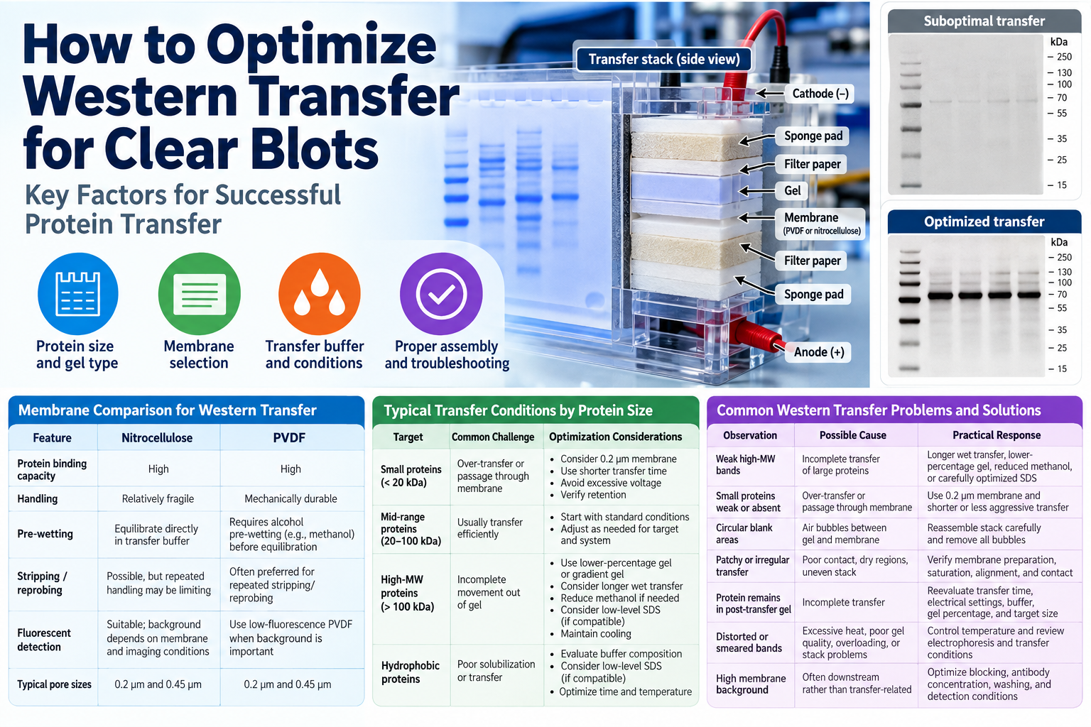
A Western blot can look excellent after electrophoresis yet fail during protein transfer. Faint bands, uneven signal, poor recovery of high-molecular-weight proteins, and loss of small proteins can all originate between the gel and membrane. Knowing how to optimize Western transfer means treating transfer as a protein-, gel-, membrane-, and apparatus-specific process rather than relying on a single timer or voltage setting.
The objective is not simply to move protein out of the gel. Effective Western transfer should preserve the relative abundance and resolution established during electrophoresis while immobilizing sufficient target protein on the membrane for reliable antibody detection.
Transfer conditions should therefore be selected according to protein molecular weight, gel composition, membrane chemistry and pore size, transfer buffer, and the type of transfer system being used.
Start With the Protein and Gel
Protein molecular weight is one of the most important variables in Western transfer optimization.
Small proteins generally transfer rapidly and may be lost through the membrane or over-transferred under aggressive conditions. Large proteins migrate more slowly through the gel matrix and may remain partially trapped in the gel under conditions that efficiently transfer smaller proteins.
A single transfer program therefore may not perform equally well for proteins ranging from approximately 10 kDa to more than 200 kDa.
Gel percentage also matters because acrylamide concentration influences pore size. Higher-percentage gels provide better separation of lower-molecular-weight proteins but can impede transfer of larger proteins. Lower-percentage gels facilitate movement of high-molecular-weight proteins but may provide less resolution for small targets.
Gradient gels are useful when samples contain proteins across a broad molecular-weight range, although transfer conditions may still need to be optimized for the specific target of interest.
Before changing transfer parameters, confirm that the target protein is appropriately resolved in the gel. Poor electrophoretic resolution or severe sample overloading cannot be corrected simply by increasing transfer time.
A prestained molecular-weight marker is useful for monitoring transfer progression, but marker behavior should not be assumed to exactly represent transfer of every endogenous, modified, membrane-associated, or highly hydrophobic protein.
Choose the Membrane for the Target
Nitrocellulose and polyvinylidene fluoride (PVDF) membranes are both widely used for research Western blotting, but membrane selection can influence protein retention, background, handling, and downstream detection.
| Membrane Feature | Nitrocellulose | PVDF |
|---|---|---|
| Protein binding | High | High |
| Handling | Relatively fragile | Mechanically durable |
Ore-wetting
| Usually equilibrated directly in transfer buffer | Conventional hydrophobic PVDF generally requires alcohol pre-wetting according to manufacturer instructions | |
| Stripping/reprobing | Possible, but repeated handling may be limiting | Often preferred when repeated stripping/reprobing is anticipated |
| Fluorescent detection | Suitable; background depends on membrane and imaging conditions | Use low-fluorescence PVDF when fluorescence background is important |
| Typical pore sizes | 0.2 µm and 0.45 µm | 0.2 µm and 0.45 µm |
A 0.45 µm membrane is commonly used for many routine Western blot targets. A 0.2 µm membrane can improve retention of small proteins that might otherwise pass through a larger-pore membrane.
Membrane selection should therefore reflect both target size and the downstream detection method rather than being treated as a fixed laboratory preference.
When using conventional PVDF, follow the membrane manufacturer’s wetting instructions. Incomplete wetting can produce localized areas of poor transfer and irregular signal.
Build the Transfer Stack Carefully
Many apparent biochemical transfer failures are actually mechanical.
The gel and membrane must maintain uniform contact across their entire surfaces. Air bubbles create localized barriers to protein migration and commonly appear as blank circular areas, distorted bands, or sharply defined regions with little or no signal.
After assembling the transfer stack, gently remove trapped bubbles using a clean roller, pipette, or other suitable tool.
For conventional electrophoretic transfer, orient the stack so that negatively charged SDS-associated proteins migrate from the gel toward the membrane and the positive electrode.
Confirm electrode orientation whenever using an unfamiliar cassette or transfer apparatus.
Filter papers and pads should be uniformly saturated with transfer buffer and free of folds or creases. The gel and membrane should also remain properly aligned.
Uneven contact can cause lane-to-lane or region-to-region variability, while excessive compression may distort the gel or interfere with uniform transfer.
How to Optimize Western Transfer Buffer
Tris-glycine transfer buffer remains a practical starting point for many Western blot workflows.
Methanol is commonly incorporated into traditional transfer buffers. It can support protein binding to membranes and influences the behavior of SDS-protein complexes and the polyacrylamide gel. However, methanol can also promote gel contraction and may reduce transfer efficiency for some high-molecular-weight proteins.
For difficult high-molecular-weight targets, reducing the methanol concentration may improve protein movement out of the gel. In some transfer systems, a low concentration of SDS can also improve transfer of large or hydrophobic proteins.
These modifications require careful optimization because SDS can reduce protein binding to the membrane and may increase the risk of protein passing through or failing to bind efficiently.
For small proteins, stronger transfer conditions are not necessarily better. Shorter transfer duration, appropriate methanol-containing buffer, and a 0.2 µm membrane may improve retention.
The exact buffer composition should always be compatible with the membrane and transfer apparatus being used.
General Starting Considerations by Protein Size
| Target | Common Transfer Challenge | Optimization Considerations |
|---|---|---|
| Small proteins (<20 kDa) | Over-transfer or passage through the membrane | Consider a 0.2 µm membrane, avoid unnecessarily long transfer, and verify protein retention on the membrane |
| Mid-range proteins (20–100 kDa) | Usually transfer efficiently under standard conditions | Begin with the transfer-system manufacturer’s recommended conditions and optimize only as needed |
| High-MW proteins (>100 kDa) | Incomplete movement out of the gel | Consider a lower-percentage or gradient gel, longer wet transfer, reduced methanol where appropriate, and carefully optimized low-level SDS if compatible |
| Hydrophobic proteins | Poor solubilization or inefficient transfer | Evaluate transfer-buffer composition and consider carefully optimized low-level SDS supplementation when compatible with the membrane and transfer system |
These are optimization principles rather than universal settings. Manufacturer recommendations for the specific gel, membrane, buffer, and transfer apparatus should remain the starting point.
Control Transfer Temperature
Transfer generates heat, particularly during prolonged wet transfer or high-current conditions.
Excessive temperature can affect band quality, transfer consistency, gel integrity, and buffer performance.
For extended transfer runs, use adequate buffer volume and follow the cooling recommendations for the transfer system. Depending on the apparatus, this may include prechilled buffer, an ice pack, a cooling module, or operation in a cold environment.
Avoid simply increasing voltage or current to accelerate a difficult transfer. Faster transfer is useful only if protein recovery and band quality remain reproducible.
Select Time and Electrical Settings by Transfer Method
Wet Transfer
Wet or tank transfer remains highly adaptable and is particularly useful when optimizing difficult or high-molecular-weight proteins.
Advantages include:
- Flexible transfer time
- Good cooling capability
- Compatibility with extended transfer conditions
- Broad applicability across protein sizes
The trade-offs are greater buffer consumption and longer setup and transfer times.
Semi-Dry Transfer
Semi-dry systems generally use less buffer and can provide rapid, convenient transfer.
They are useful for routine Western blotting, but transfer performance depends on the specific apparatus, buffer system, stack configuration, and target molecular weight. The stack must remain uniformly hydrated throughout the transfer.
Modern semi-dry systems may use proprietary buffers and transfer programs, so the manufacturer’s operating instructions should be followed before modifying voltage, current, or transfer duration.
Optimize One Variable at a Time
For a new or difficult target, begin with the transfer-system manufacturer’s recommended conditions and then change one variable at a time.
Possible variables include:
- Transfer time
- Voltage or current
- Methanol concentration
- SDS concentration
- Membrane pore size
- Gel percentage
- Transfer temperature
Changing several parameters simultaneously makes it difficult to determine which modification improved or worsened transfer.
Use equivalent sample aliquots when comparing conditions and evaluate the resulting blots under comparable detection and exposure settings.
Confirm That Transfer Actually Occurred
Poor Western blot signal is not always caused by transfer.
After transfer, inspect the membrane and, when troubleshooting, consider staining the membrane with an appropriate reversible total-protein stain. The post-transfer gel can also be stained to determine whether substantial protein remains in the gel.
These checks help distinguish incomplete transfer from downstream problems involving blocking, antibody binding, washing, or detection.
For antibody-related troubleshooting, see Antibody 101.
Troubleshoot the Transfer Pattern, Not Just Signal Intensity
The pattern of the problem often provides more information than overall signal strength.
| Problem / Observation | Likely Cause | Recommended Action |
|---|---|---|
| Weak high-MW protein bands | Incomplete transfer of large proteins | Consider longer wet transfer, lower-percentage or gradient gel, reduced methanol, and carefully optimized low-level SDS if compatible |
| Small proteins weak or absent | Over-transfer or passage through membrane | Use a 0.2 µm membrane and reduce transfer time or transfer intensity |
| Protein remains in post-transfer gel | Incomplete transfer | Increase transfer time as appropriate; review voltage/current, buffer composition, gel percentage, and target molecular weight |
| Circular blank areas on membrane | Air bubbles trapped between gel and membrane | Reassemble the transfer stack and carefully remove all bubbles before transfer |
| Patchy or uneven transfer | Poor gel-membrane contact, dry regions, uneven stack, or incomplete membrane preparation | Ensure uniform wetting, correct stack alignment, full buffer saturation, and even contact |
| Bands appear distorted or smeared | Excessive heating, sample overloading, poor electrophoretic separation, or transfer-stack problems | Control transfer temperature; review sample loading, electrophoresis, and stack assembly |
| Transfer strong at one side but weak at the other | Uneven stack contact, trapped air, inadequate buffer distribution, or cassette assembly problem | Reassemble the stack, verify uniform saturation and pressure, and check cassette orientation |
| Very faint signal across entire membrane | Incomplete transfer, insufficient sample, low target abundance, or antibody/detection problem | Examine the post-transfer gel and total-protein staining before changing antibody conditions |
| High membrane background | Often related to blocking, antibody concentration, washing, or detection rather than transfer | Optimize blocking, antibody dilution, washing, and detection conditions after confirming successful transfer |
| Small proteins transfer well but large proteins do not | Conditions favor rapid transfer of lower-MW proteins | Optimize specifically for high-MW targets using longer transfer, appropriate gel percentage, reduced methanol, and controlled temperature |
Uniformly faint bands can result from incomplete transfer, insufficient sample loading, low target abundance, or poor antibody performance. Examining the post-transfer gel and membrane protein pattern can help identify where the problem originated.
Special Considerations for High-Molecular-Weight Proteins
Large proteins frequently require more deliberate optimization.
Potential approaches include:
- Use a lower-percentage or appropriate gradient gel
- Favor wet transfer when extended transfer is needed
- Reduce methanol concentration when appropriate
- Evaluate carefully controlled low concentrations of SDS if compatible with the membrane and system
- Maintain adequate cooling during prolonged transfer
- Confirm residual protein in the post-transfer gel
Avoid assuming that simply increasing voltage is the best solution. Excess current can increase heating without proportionally improving recovery of large proteins.
Special Considerations for Small Proteins
Small proteins present the opposite problem: they may transfer efficiently but fail to remain on the membrane.
For low-molecular-weight targets:
- Consider a 0.2 µm membrane
- Avoid unnecessarily prolonged transfer
- Avoid SDS supplementation unless specifically required and validated
- Verify membrane retention using an appropriate staining method
- Optimize transfer time rather than automatically using conditions developed for larger proteins
Standardize the Workflow After Optimization
Once transfer conditions are working reliably, document the complete procedure.
Record:
- Target molecular weight
- Gel type and acrylamide percentage
- Membrane material
- Membrane pore size
- Transfer buffer formulation
- Methanol concentration
- SDS concentration, if used
- Transfer apparatus
- Voltage or current
- Transfer duration
- Temperature-control method
- Sample load
- Membrane staining or transfer-quality assessment
These details improve reproducibility, particularly when multiple researchers perform the assay or when a method is transferred between laboratories.
For projects involving proteins across very different molecular-weight ranges, separate validated transfer conditions for small, standard, and high-molecular-weight targets may provide more reproducible results than forcing every experiment into one universal protocol.
A well-optimized Western transfer should produce uniform protein immobilization across the membrane while minimizing residual target in the gel and preventing loss through the membrane. Building simple transfer-quality checks into each run turns Western transfer from a recurring source of variability into a controlled and reproducible step in protein research.
References
- Thermo Fisher Scientific. Western Blotting Handbook. Guidance on Western blot transfer, membranes, transfer systems, detection, and troubleshooting. Thermo Fisher Scientific
- Thermo Fisher Scientific. Western Blotting Support — Getting Started. Technical guidance for Western blotting and protein transfer systems. Thermo Fisher Scientific
- Thermo Fisher Scientific. Western Blotting Support — Troubleshooting. Guidance on high-molecular-weight protein transfer, SDS and methanol considerations, transfer time, gel percentage, and common transfer problems. Thermo Fisher Scientific
- Abcam. Western Blot Protein Transfer and Visualization. Guidance on wet and semi-dry transfer, membrane handling, transfer verification, and optimization. Abcam
- Mishra M, Tiwari S, Gomes AV. Protein purification and analysis: next generation Western blotting techniques. Expert Review of Proteomics. 2017;14(11):1037–1053. DOI: 10.1080/14789450.2017.1388167. PubMed
For Research Use Only.