


Caspase Activity Assay Selection and Controls


Apoptosis experiments often fail at the interpretation stage rather than the treatment stage. A caspase activity assay can show that proteases are enzymatically active, but only when the assay format, substrate specificity, sample preparation, and controls match the biological question. Used thoughtfully, it provides a functional readout that complements viability measurements, morphology, Western blotting, and other apoptosis markers.
What a Caspase Activity Assay Measures
Caspases are cysteine-dependent aspartate-directed proteases that participate in programmed cell death, inflammation, and related signaling pathways. In apoptosis research, assays commonly measure the activity of initiator caspases, such as caspase-8 or caspase-9, and executioner caspases, particularly caspase-3 and caspase-7.
Most caspase activity assays use a short peptide substrate containing a preferred cleavage sequence. The substrate is linked to a fluorescent, luminescent, or colorimetric reporter. When an active caspase cleaves the peptide, the reporter generates a measurable signal. Higher signal generally indicates greater cleavage activity under the defined assay conditions.
This distinction matters: the assay measures enzymatic cleavage of an artificial substrate, not necessarily the abundance of a specific caspase protein. A sample can contain procaspase-3 without detectable catalytic activity, while a sample with modest protein abundance may produce a strong activity signal after activation. For this reason, activity data are often strongest when paired with immunoblot detection of cleaved caspase or cleaved PARP.
Choose the Assay Format Around the Experimental Question
The best caspase activity assay depends on throughput, sample type, expected signal level, and whether cells need to remain intact before measurement. The three commonly used readouts each have practical advantages.
Colorimetric Assays
Colorimetric substrates are typically linked to p-nitroaniline or a related chromophore. Cleavage produces a color change measured by absorbance, often near 405 nm. These methods are familiar, cost-conscious, and useful when a plate reader has absorbance capability but lacks luminescence detection.
Their trade-off is sensitivity. Colorimetric assays may be less suitable for low cell numbers, weakly induced apoptosis, or samples with background absorbance from media components, colored compounds, or lysate contaminants. They are generally most useful when sufficient lysate can be prepared and changes in activity are expected to be substantial.
Fluorometric Assays
Fluorometric formats commonly use AFC- or AMC-labeled peptide substrates. Enzymatic cleavage releases a fluorescent product, allowing higher sensitivity than many absorbance-based methods. They are appropriate for cell lysates, tissue extracts, and plate-based studies requiring a broad dynamic range.
Fluorescent assays require attention to compound interference. Test compounds, endogenous pigments, and some culture media can fluoresce or quench fluorescence. Run treatment-only wells without substrate when screening unfamiliar compounds, especially in small-molecule or natural-product studies.
Luminescent Assays
Luminescent caspase assays are often selected for low sample input, microplate workflows, and high-throughput screening. Because luminescence has low background, these assays can detect activity from relatively few cells and are compatible with many cell-based experimental designs.
However, luminescent signal is not automatically equivalent to cell number or total protein. Treatments that reduce viable cell mass can complicate comparisons. For adherent cultures, consider whether the workflow measures cells remaining in the well, detached apoptotic cells, or the combined population. Collecting both floating and attached cells is often necessary to avoid underestimating apoptosis.
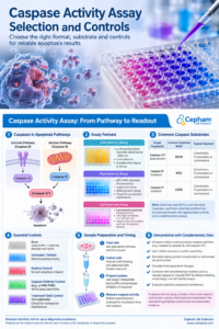
Figure 1. Overview of caspase activity assay formats, apoptosis pathways, commonly used peptide substrates, and essential experimental controls.
Match Substrate Specificity to the Pathway Under Study
Peptide substrates are commonly described by cleavage motifs. DEVD-based substrates are widely used for caspase-3/7 activity, IETD-based substrates are associated with caspase-8, and LEHD-based substrates are commonly used for caspase-9. These designations are useful for assay planning, but they should not be treated as absolute proof of activity from one enzyme alone.
Caspases can share substrate preferences, particularly in complex lysates where multiple proteases may be active. A DEVD signal supports executioner caspase activity, but it does not always distinguish caspase-3 from caspase-7. Similarly, an apparent initiator caspase signal should be interpreted with pathway context, inhibitor controls, and orthogonal evidence.
When the goal is to establish intrinsic versus extrinsic apoptosis, use a panel approach. Measure an executioner caspase readout alongside caspase-8 and caspase-9 activity, then compare the results with mitochondrial markers, death receptor pathway markers, or cleavage-specific immunoblot data. No single substrate assay fully maps an apoptotic pathway.
Build Controls Into Every Caspase Activity Assay
Controls establish whether signal changes reflect caspase activation, technical variation, or assay interference. At minimum, include untreated cells and a positive apoptosis control known to work in the selected cell model. The specific positive control may vary by cell type and experimental design, so preliminary optimization is appropriate.
For a reliable interpretation, include these controls where applicable:
-
- Blank control: Assay buffer and substrate, used to establish background signal.
-
- Untreated or vehicle control: Defines baseline caspase activity.
-
- Positive apoptosis control: Confirms that the assay can detect induced caspase activity.
-
- Caspase inhibitor control: A broad-spectrum or pathway-relevant inhibitor used to test signal specificity.
-
- Interference control: Treatment-only wells lacking substrate for fluorescent or colorimetric assays when compound interference is possible.
The inhibitor control is particularly informative. A meaningful decrease in assay signal after inhibitor exposure supports caspase-dependent substrate cleavage. It does not prove complete selectivity, since inhibitor performance depends on concentration, permeability, timing, and the enzymes present in the sample.
Sample Preparation Determines Signal Quality
For lysate-based methods, preserve enzymatic activity from the start. Follow the assay manufacturer’s instructions for collection temperature and handling, use the lysis buffer recommended for the assay chemistry, and process lysates promptly. Repeated freeze-thaw cycles can reduce activity and increase variability.
Avoid assuming that a standard protein extraction buffer is automatically suitable for caspase assays. Strong detergents, incompatible salts, reducing agents, or protease inhibitor cocktails may affect enzyme activity or interfere with detection. General protease inhibitors can also suppress the biological activity being measured. If using a custom lysis workflow, verify compatibility with the selected substrate and reporter system before analyzing a full study.
Where appropriate for a validated lysate-based assay, normalize lysate input by total protein when comparing samples with different cell densities or growth rates. A BCA or comparable protein quantitation method can support this step, provided the lysis buffer is compatible with the protein assay.
When appropriate, report background-corrected activity relative to protein input, and specify the assay’s normalization method. Do not assume that all luminescent or intact-cell formats support this approach.
For intact-cell luminescent workflows, normalization may instead use matched parallel cell counts, DNA content, or a compatible viability measurement. The correct approach depends on whether the biological question concerns total caspase activity per well or caspase activation relative to the number of cells present.
Timing Is a Biological Variable, Not a Technical Detail
Caspase activation is often transient. An early time point may miss activation before the pathway has progressed, while a late time point may produce lower signal because apoptotic cells have detached, lysed, or advanced to secondary necrosis.
The peak may occur within hours or substantially later, depending on the cell model, stimulus, dose, and assay conditions. A single endpoint can miss activation.
For a new model, run a limited time course before committing to a large experiment. Sampling several intervals across the anticipated response window is more informative than choosing one convenient endpoint. This is especially relevant when comparing fast-acting cytotoxic compounds with slower genetic perturbations or nutrient-stress conditions.
Common Reasons for Weak or Inconsistent Results
Low signal can result from insufficient apoptosis induction, incorrect sampling time, low cell input, loss of detached cells, inactive substrate, or poor lysate handling.
Confirm the positive control first. If it performs as expected while experimental samples remain low, the treatment conditions or cell model may not be producing caspase-dependent apoptosis.
High background may reflect substrate degradation, reader settings, contamination, fluorescent test compounds, or excessive lysate concentration.
A dilution series can reveal whether the signal is within the assay’s linear range. For fluorometric and colorimetric assays, confirm that the plate type and instrument filters or wavelengths are appropriate for the reporter.
A strong decrease in viability without increased caspase activity also deserves careful interpretation. The cells may be dying through a caspase-independent process, the activity peak may have been missed, or the assay may not be compatible with the sample preparation.
Pairing the activity assay with cell viability data, membrane-integrity measurements, and protein-based apoptosis markers helps resolve these possibilities.
Integrate Activity Data With the Rest of the Workflow
A caspase activity assay is most useful as part of a defined apoptosis workflow rather than as a stand-alone endpoint.
In many studies, initial viability screening identifies active treatment ranges, caspase measurements establish whether enzymatic apoptosis is involved, and Western blotting or immunoassays confirm pathway-associated cleavage events.
Flow cytometry-based annexin V and membrane-impermanent dye staining can further distinguish early apoptotic, late apoptotic, and necrotic populations.
For research laboratories selecting assay reagents, prioritize the readout chemistry, substrate target, detection instrument compatibility, sample-input requirements, and required controls before comparing formats.
Cepham Life Sciences supports apoptosis and cell viability workflows with research-use reagents and laboratory products intended to fit these practical experimental requirements.
A well-controlled caspase assay does more than produce a higher signal in treated cells. It provides evidence that can be connected to timing, pathway biology, and sample quality, allowing researchers to make a more defensible decision about how cell death is occurring.
References
-
- Promega. Caspase-Glo® 3/7 Assay Technical Bulletin (TB323). Technical guidance on DEVD-based luminescent caspase-3/7 measurements, cell lysis, and assay performance.
- Thermo Fisher Scientific. Apoptosis Protocols. Caspase assay formats and complementary apoptosis measurements.
- Promega. Caspase-Glo® 3/7 3D Assay Technical Manual (TM627). Guidance on luminescent assay conditions, interference, and three-dimensional cultures.
For Research Use Only
This article is provided for research and educational purposes. Assay selection, reagent compatibility, sample preparation, controls, normalization, and interpretation should follow the manufacturer’s instructions and validation appropriate to the experimental system. Products and workflows discussed are for research use only and are not intended for human administration, therapeutic use, or clinical diagnostic applications unless specifically authorized for those purposes.