


IHC Antigen Retrieval: Choosing the Right Method

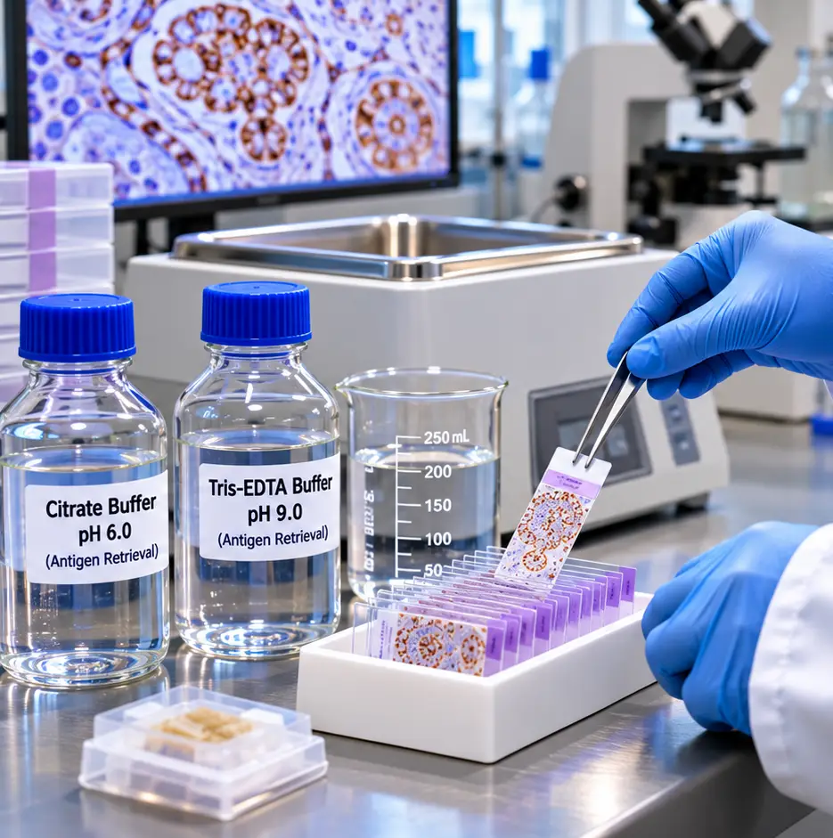
Formalin fixation preserves tissue architecture, but it can also mask epitopes needed for antibody binding. IHC antigen retrieval is a controlled pretreatment used to improve epitope accessibility before immunohistochemical staining. When the method is matched to the tissue, antigen, antibody clone, and detection system, retrieval can improve staining intensity and localization. Poorly selected conditions may instead cause weak staining, nonspecific background, tissue detachment, or distorted morphology.
Antigen retrieval is an experimental variable, not a universal step. Conditions that work for one formalin-fixed, paraffin-embedded (FFPE) specimen may not suit another antibody or tissue. Reliable optimization starts with a documented protocol, relevant controls, and changes to one variable at a time.
Why Fixation Masks Antigens in IHC
Formaldehyde fixation creates chemical modifications and cross-links that stabilize tissue structures but can alter protein conformation, conceal antibody-binding sites, or restrict reagent access. The degree of masking depends on fixation time, tissue thickness, processing conditions, antigen abundance, and the epitope recognized by the primary antibody.
Long fixation may increase masking, but stronger retrieval is not automatically better. Excessive heating or proteolysis can damage tissue, remove target antigen, or expose unwanted binding sites. The objective is specific staining with expected localization, acceptable morphology, and low background, rather than the darkest possible stain.
Fresh-frozen tissue may need little or no retrieval, particularly when it has not undergone extensive aldehyde fixation. Post-fixed frozen sections can behave differently. Follow application-specific antibody validation information and optimize locally when necessary.
IHC Antigen Retrieval Methods: HIER and PIER
Two widely used approaches are heat-induced epitope retrieval (HIER) and proteolytic-induced epitope retrieval (PIER). A no-retrieval condition can also be valuable during initial screening, especially for fragile specimens or readily accessible epitopes.
Heat-Induced Epitope Retrieval (HIER)
HIER heats hydrated sections in a defined retrieval solution to improve accessibility of formalin-masked epitopes. It is widely used for FFPE IHC and can be standardized across staining batches.
Common platforms include pressure-based retrieval chambers, steamers, water baths, microwaves, and dedicated automated instruments. They differ in heating rate, temperature uniformity, and cooldown behavior. A protocol transferred between devices should be rechecked rather than assumed equivalent.
Citrate buffer near pH 6.0 is a common starting point. EDTA near pH 8.0 and Tris-EDTA near pH 9.0 are useful alternatives for some targets. Buffer formulation, ionic strength, heating profile, and section adhesion also influence performance. The optimal buffer must be determined for the antibody and specimen.
Proteolytic-Induced Epitope Retrieval (PIER)
PIER uses controlled enzymatic digestion, often with proteinase K, trypsin, pepsin, or pronase, to improve antibody access. It may be useful for particular targets or when validated heat-based methods do not produce suitable staining.
Enzymatic retrieval requires careful control of enzyme activity, concentration, temperature, and incubation time. Overdigestion can cause loss of cellular detail, section lifting, diffuse staining, or destruction of the epitope itself. Some protocols combine heat and enzymatic treatments, but combined methods require separate validation.
HIER Versus PIER: Practical Comparison
| Feature | Heat-Induced Epitope Retrieval (HIER) | Proteolytic-Induced Epitope Retrieval (PIER) |
|---|---|---|
| Main principle | Heat in aqueous buffer improves epitope accessibility | Controlled enzyme digestion improves antibody access |
| Common reagents | Citrate pH ~6; EDTA pH ~8; Tris-EDTA pH ~9 | Proteinase K, trypsin, pepsin, or pronase |
| Key variables | Buffer, pH, temperature, heating time, cooldown | Enzyme type, concentration, incubation time, temperature |
| Main advantages | Broad utility for FFPE tissues; readily standardized | Useful for selected epitopes that respond to digestion |
| Main limitations | Excessive heat can damage tissue or increase background | Overdigestion can destroy morphology or antigen |
| Starting approach | Compare antibody-recommended conditions and a limited buffer screen | Use when supported by validation or after controlled HIER screening |
Table 1. Comparison of common IHC antigen retrieval approaches. These are general principles, not universal operating conditions; follow antibody and reagent instructions.
Selecting the Best IHC Antigen Retrieval Conditions
Start with the antibody manufacturer’s recommendation for IHC on FFPE sections when available. A protocol validated for Western blotting or ELISA cannot be assumed to work for tissue immunostaining.
For an uncharacterized antibody–tissue combination, compare matched sections using no retrieval, citrate-based HIER, and alkaline EDTA- or Tris-EDTA-based HIER. Include appropriate positive tissue and no-primary-antibody controls. If these conditions fail despite a verified positive control, consider a limited enzymatic screen where biologically and technically justified.
Evaluate staining location, signal-to-background contrast, and tissue preservation together. A stronger signal is not an improvement if localization becomes inconsistent with the expected biology.
Record the Complete Retrieval Protocol
Document buffer identity and pH, device type, actual or programmed temperature, time at temperature, cooldown, slide coating, section thickness, fixation history when known, and staining-run conditions. This makes later troubleshooting and method transfer more reproducible.
Variables That Affect Antigen Retrieval Performance
Temperature and duration: A long, moderate-temperature exposure is not necessarily equivalent to a shorter high-temperature cycle. Pressure systems, steamers, and water baths can produce different effective treatment profiles.
Cooldown: Controlled cooling is part of the method and should be consistent. Abrupt handling of hot slides can contribute to section loss or inconsistent staining.
Slide adhesion and preparation: Use slides appropriate for the retrieval conditions, dry sections according to the protocol, and ensure complete deparaffinization and rehydration before aqueous retrieval.
Detection and blocking: Once a reasonable retrieval condition is established, optimize antibody dilution, endogenous enzyme blocking, protein blocking, incubation, and detection chemistry separately. Simultaneous changes make it difficult to identify the cause of staining differences.
Troubleshooting Common IHC Antigen Retrieval Problems
| Observation | Possible Causes | First Checks or Adjustments |
|---|---|---|
| Weak or absent specific staining | Insufficient retrieval, unsuitable buffer, low antigen expression, fixation effects, antibody issues | Verify positive control; compare retrieval buffers; confirm antibody application |
| Strong background or diffuse staining | Over-retrieval, concentrated antibody, inadequate blocking, drying, detection-related signal | Examine no-primary control; reduce retrieval severity; reassess antibody dilution and blocking |
| Tissue lifting or loss | Inadequate slide adhesion, insufficient drying, harsh heat or enzyme treatment | Use appropriate adhesive slides; standardize drying and cooling; reduce treatment intensity |
| Uneven staining | Incomplete dewaxing, uneven heating, evaporation, poor reagent coverage | Check rehydration, buffer volume, instrument uniformity, and section coverage |
Table 2. Troubleshooting guide for research IHC. Interpret each finding with control slides and expected staining localization.
Weak or Absent Staining
Before intensifying retrieval, confirm that the positive control tissue is appropriate and expresses the target. Check antibody compatibility with FFPE IHC, fixation history, and detection chemistry. Then compare a different retrieval buffer or carefully adjusted heating condition.
High Background or Diffuse Signal
Background can arise from excessive retrieval, antibody concentration, inadequate blocking, drying artifacts, or endogenous detection-system activity. Use a no-primary control to assess detection-related signal and compare the observed pattern with expected tissue or subcellular localization.
Tissue Detachment and Uneven Staining
For section loss, verify slide adhesion and drying before increasing treatment severity. For uneven staining, check dewaxing, reagent coverage, buffer volume, heating uniformity, and evaporation. Avoid overcrowding slides in the retrieval vessel.
Controls for Reliable IHC Antigen Retrieval
A known positive tissue control helps establish that the staining system can detect the target. A negative tissue control, when biologically appropriate, helps evaluate specificity. A no-primary-antibody control can reveal staining from secondary reagents, detection chemistry, or endogenous activity. An isotype control may answer certain questions but is not a substitute for a biologically meaningful negative control or validated antibody specificity.
Whenever feasible, process controls alongside experimental sections under matched fixation, retrieval, and staining conditions. A positive control cannot completely account for differences in the fixation history or composition of test tissue, but it provides essential context when interpreting weak or negative staining.
For reproducible research, report the retrieval method, buffer, pH, instrument, heating or digestion conditions, cooldown, and controls in the experimental methods.
Conclusion
Successful IHC antigen retrieval balances improved epitope accessibility with tissue preservation and staining specificity. Begin with antibody-supported conditions, compare a limited HIER screen when needed, and use PIER selectively. Interpret results using positive and negative controls, expected localization, and morphology—not staining intensity alone.
Careful documentation and measured optimization help researchers obtain interpretable, reproducible immunohistochemistry results across sections and experimental runs.
References
- Shi SR, Shi Y, Taylor CR. Antigen retrieval immunohistochemistry: review and future prospects in research and diagnosis over two decades. Journal of Histochemistry & Cytochemistry. 2011;59(1):13–32. https://doi.org/10.1369/jhc.2010.957191
- Yamashita S, Katsumata O. Heat-Induced Antigen Retrieval in Immunohistochemistry: Mechanisms and Applications. Methods in Molecular Biology. 2017;1560:147–161. https://doi.org/10.1007/978-1-4939-6788-9_10
- Shi SR, Shi Y, Taylor CR, Gu J. New Dimensions of Antigen Retrieval Technique: 28 Years of Development, Practice, and Expansion. Applied Immunohistochemistry & Molecular Morphology. 2019;27(10):715–721. https://doi.org/10.1097/PAI.0000000000000778
For Research Use Only
This article is intended for research and educational purposes. Antigen retrieval conditions and immunohistochemistry procedures must be optimized and validated for the intended specimen, antibody, detection system, and laboratory application. Research-use reagents and workflows described here are not intended for clinical diagnostic, therapeutic, or human-use applications unless specifically authorized for those purposes.