


DNA Ladder Selection for Gel Electrophoresis

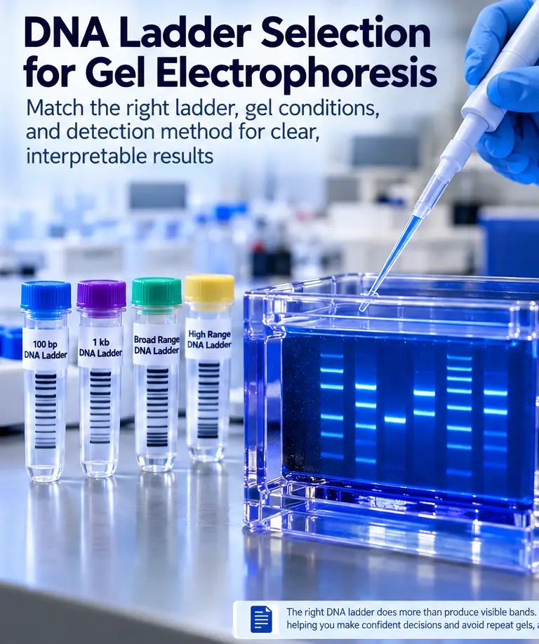
A DNA ladder is the reference scale that makes gel electrophoresis results interpretable. Effective DNA ladder selection starts with the expected size range of experimental fragments, then accounts for gel chemistry, separation method, stain sensitivity, and the level of sizing precision required. A poorly matched ladder may still produce visible bands, but it can make fragment assignment uncertain at the sizes that matter most.
For routine PCR confirmation, a broad-range ladder may be sufficient. For cloning workflows, restriction digest verification, or analysis of short amplicons, tighter band spacing around the expected fragment size is usually more useful. The goal is not to select the ladder with the greatest number of bands, but to choose a molecular size reference that resolves the experimental question.
DNA Ladder Selection Begins With Expected Fragment Size
The first consideration is the expected DNA size or size range. DNA ladders are commonly classified as low-range, 100 bp, 1 kb, broad-range, and high-molecular-weight markers. These names provide useful guidance, but the manufacturer’s datasheet should determine the final choice.
Review the exact band sizes, total molecular weight range, DNA mass per band, and any intensified reference bands.
100 bp DNA Ladders
A 100 bp DNA ladder is commonly used for PCR products and smaller restriction fragments. Many provide closely spaced bands across approximately 100–1,000 bp, although actual ranges vary by product.
These markers are particularly useful when distinguishing fragments of similar size, such as 250 bp and 300 bp, or confirming the presence of a short DNA insert.
1 kb DNA Ladders
A 1 kb ladder is generally suitable for longer PCR products, plasmid inserts, and restriction digest fragments extending through several kilobases.
Broad-range ladders provide additional flexibility when analyzing samples containing both small and large fragments. However, wider intervals between neighboring bands may reduce sizing precision near the region of interest.
High-Molecular-Weight DNA Ladders
For fragments larger than approximately 10 kb, consider a ladder designed for high-molecular-weight DNA.
Standard agarose electrophoresis may not adequately resolve very large fragments. Depending on the required resolution, lower agarose concentrations, specialized gel matrices, longer runs, or pulsed-field gel electrophoresis may be necessary.
The appropriate system depends on DNA size, sample integrity, and the degree of separation required.
Match Reference Bands to the Expected Fragment
The most useful ladder bands should bracket the expected sample size.
For example, when analyzing a 650 bp amplicon, reference bands at 600 bp and 700 bp provide a more informative comparison than bands at 500 bp and 1,000 bp.
Many commercial ladders include one or more intensified bands, such as a 500 bp or 1 kb reference. These help researchers identify the band pattern quickly but do not independently improve sizing accuracy.
DNA Ladder Types and Common Applications
| Ladder Type | Typical Application | Main Selection Consideration |
|---|---|---|
| Low-range ladder | Short PCR products and small DNA fragments | Dense spacing at low base-pair sizes |
| 100 bp ladder | Routine PCR and small restriction fragments | Clear separation near expected amplicons |
| 1 kb ladder | Longer PCR products and plasmid digests | Coverage across several kilobases |
| Broad-range ladder | Mixed-size samples and initial screening | Sufficient reference bands near important fragments |
| High-range ladder | Large DNA constructs and genomic DNA analysis | Compatibility with high-molecular-weight separation |
Table 1. Common DNA ladder categories and selection considerations. Exact fragment ranges depend on the manufacturer’s formulation.
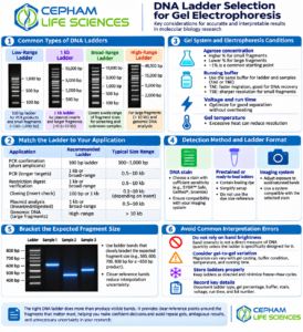
Figure 1. DNA ladder types, application matching, electrophoresis conditions, detection formats, and common interpretation errors. Illustrative graphic; consult the manufacturer’s ladder datasheet for exact band sizes.
Consider the Gel System and Electrophoresis Conditions
A DNA ladder performs only as well as the electrophoresis system allows.
Agarose concentration strongly influences fragment separation. Higher-percentage gels generally provide improved resolution for smaller DNA fragments, while lower-percentage gels are more suitable for larger fragments.
A concentration near 1% agarose is often a practical starting point for routine DNA analysis, although the optimal concentration depends on the fragment range, agarose formulation, and running conditions.
If fragments of interest migrate too closely together, changing the ladder alone will not improve separation. Gel concentration, voltage, buffer composition, and run duration may also require optimization.
TAE Versus TBE Running Buffer
TAE and TBE are widely used electrophoresis buffers.
TAE is frequently selected for general-purpose DNA electrophoresis and workflows involving subsequent DNA recovery. TBE offers greater buffering capacity and may provide improved resolution of smaller fragments under suitable conditions.
Both can support routine DNA analysis, but migration behavior depends on the complete electrophoresis system.
Use the manufacturer’s recommended buffer conditions and ensure the ladder and samples are separated under the same running conditions.
Loading Volume and DNA Mass
Excessive ladder loading can cause broad, distorted, or saturated bands. Insufficient loading may produce weak bands that are difficult to visualize.
Follow the recommended loading quantity for the selected ladder, gel stain, and imaging system.
Where necessary, adjust the quantity after checking whether the strongest reference bands remain within the detector’s usable signal range.
Choose a DNA Ladder Compatible With the Detection Method
Most DNA ladders are visualized using fluorescent nucleic acid stains or other DNA detection systems.
Stain sensitivity affects the minimum detectable DNA mass per band. Highly sensitive stains can reveal low-abundance fragments, while excessive staining or loading may saturate intense bands.
The imaging instrument must also be compatible with the stain’s excitation and emission properties.
Ready-to-Load DNA Ladders
Ready-to-load ladders simplify gel preparation because they contain loading buffer and tracking dyes.
These formulations can reduce preparation steps and improve consistency during routine PCR and cloning workflows.
However, tracking dyes migrate independently of DNA fragments. Their position should not be interpreted as a molecular size reference.
Also distinguish between ladders supplied with loading dye and products containing DNA fragments labeled for direct visualization. These are not necessarily the same formulation.
Consider Downstream Applications
For gel extraction, cloning, sequencing, or other downstream molecular biology applications, select gel stains and electrophoresis reagents compatible with the intended recovery workflow.
The ladder serves primarily as a sizing reference. Nevertheless, careful reagent handling and contamination control remain important, particularly when working with sensitive amplification or sequencing methods.
DNA Ladder Selection for Common Research Applications
PCR Product Confirmation
For endpoint PCR, choose a ladder with closely spaced bands around the expected amplicon.
A 100 bp ladder is often appropriate for products below approximately 1 kb. Longer amplicons may require a 1 kb or broad-range marker.
When nonspecific amplification is possible, select a ladder covering both the expected product and plausible additional fragments.
Restriction Digest Verification
Before running a restriction digest, calculate the expected fragment sizes from the restriction map.
The selected ladder should cover all fragments relevant to interpreting the digest.
Smaller fragments can be especially important when verifying insert orientation, successful cloning, or complete restriction digestion.
Plasmid DNA Analysis
Undigested plasmid DNA can occur in supercoiled, nicked, linear, and multimeric forms. These structures migrate differently during agarose electrophoresis.
Therefore, a conventional linear DNA ladder should not be used to assign an exact molecular size to an undigested plasmid based solely on migration distance.
For more reliable size assessment, linearize the plasmid or analyze appropriately selected restriction fragments.
Genomic DNA Quality Assessment
For genomic DNA, gel electrophoresis is frequently used to evaluate integrity rather than determine an exact fragment size.
Intact high-molecular-weight DNA may remain close to the loading well, while degraded or mechanically sheared DNA can appear as a smear.
Use an appropriate high-range marker and separation system when fragment-size assessment is required.
Avoid Common DNA Ladder Interpretation Errors
DNA ladders provide approximate molecular sizing under the conditions of a particular electrophoresis run.
Migration differences may arise from uneven gel casting, buffer depletion, overheating, excessive voltage, or differences in sample salt concentration.
For wide gels, placing ladder lanes near both sides of the sample region may help identify uneven migration.
Do Not Confuse Band Brightness With DNA Quantity
Band brightness depends on several factors, including DNA mass, stain binding, detector response, exposure settings, and fragment characteristics.
Do not estimate sample DNA concentration by direct comparison with ladder brightness unless the marker is intended for quantitative use and the method has been appropriately validated.
For more accurate DNA quantification, use a suitable fluorometric or spectrophotometric assay, with attention to its limitations and sample compatibility.
Store and Handle DNA Ladders Properly
Follow the manufacturer’s storage and handling recommendations.
Avoid unnecessary freeze-thaw cycles, contamination, and prolonged exposure to unsuitable temperatures.
Record the ladder identity and lot number, loading amount, gel concentration, electrophoresis buffer, stain, voltage, and run duration when reproducibility is important.
Practical DNA Ladder Selection Checklist
Before choosing a DNA ladder, confirm:
- Expected fragment size: Does the ladder cover the full relevant range?
- Band spacing: Are reference bands sufficiently close to the expected fragment?
- Gel compatibility: Can the electrophoresis system resolve the fragments?
- Detection sensitivity: Will the selected stain and imager detect the ladder bands?
- Loading requirements: Is the DNA mass per band appropriate?
- Application: Is the marker suitable for PCR, restriction digests, plasmid analysis, or genomic DNA assessment?
- Documentation: Are exact band sizes and recommended conditions available in the product datasheet?
Conclusion
Reliable DNA ladder selection is a small but important part of successful gel electrophoresis.
The most suitable ladder is not necessarily the one with the broadest range or greatest number of bands. It is the marker that provides clear reference points around the DNA fragments that determine the next experimental decision.
Matching the ladder to fragment size, gel conditions, detection method, and application can improve interpretation, reduce unnecessary repeat experiments, and support reproducible molecular biology research.
Cepham Life Sciences supports molecular biology workflows with research-use reagents and laboratory supplies for DNA preparation, amplification, electrophoresis, and related applications.
References
- Green MR, Sambrook J. Agarose Gel Electrophoresis. Cold Spring Harbor Protocols. 2019;2019(1). DOI: 10.1101/pdb.prot100404.
- Lee PY, Costumbrado J, Hsu CY, Kim YH. Agarose Gel Electrophoresis for the Separation of DNA Fragments. Journal of Visualized Experiments. 2012;(62):3923. DOI: 10.3791/3923.
- Thermo Fisher Scientific. DNA and RNA Gel Electrophoresis Resources. Technical information on nucleic acid electrophoresis and molecular weight markers.
For Research Use Only
This article is provided for research and educational purposes. DNA ladder selection, electrophoresis conditions, detection methods, and interpretation should follow the relevant manufacturer’s instructions and validated laboratory procedures.
Products and workflows discussed are for research use only and are not intended for human administration, therapeutic use, or clinical diagnostic applications unless specifically authorized for those purposes.