


Cell Viability Assay Comparison for Research

Cell viability assays are widely used to evaluate how experimental treatments affect cultured cells, but different assays do not necessarily measure the same biological endpoint. A treatment can reduce a viability-associated signal without immediately killing cells, while a stable signal can sometimes mask substantial changes in proliferation rate or metabolic state.
That distinction is the starting point for any cell viability assay comparison. The appropriate method depends on what the experiment must measure: metabolically active cells, intracellular ATP, membrane integrity, total cell number, or cytotoxicity caused by membrane damage.
For research workflows, no assay format is universally interchangeable. Media composition, plate format, cell density, compound color, incubation duration, and the expected mechanism of injury can all influence assay performance. Selecting a readout that matches the biological question is generally more informative than selecting the fastest or most familiar assay.
What a Cell Viability Assay Actually Measures
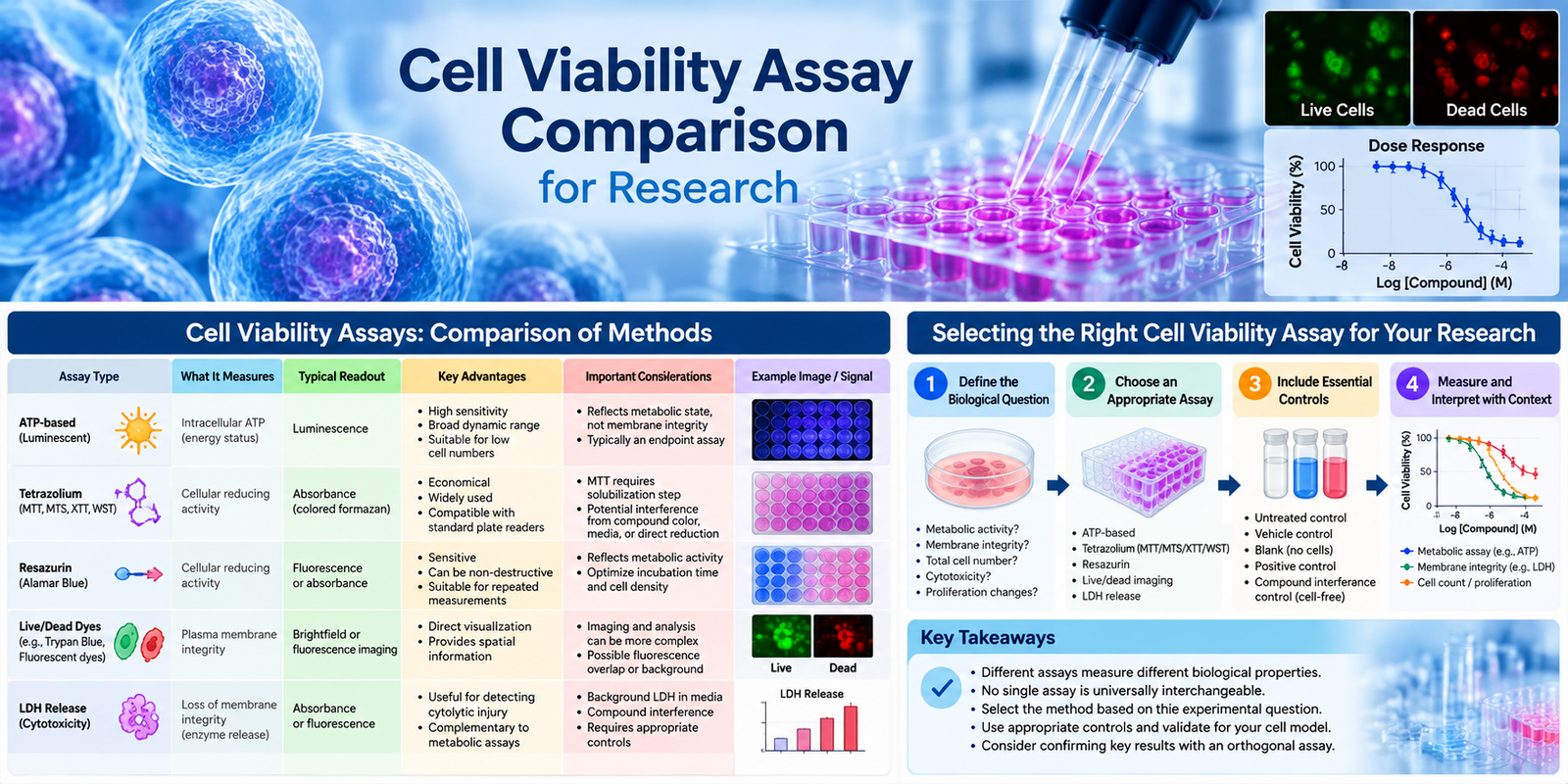
Cell viability is often used as a broad label, but commonly used assays measure different biological properties.
- ATP-based assays estimate intracellular ATP as a marker associated with viable cells.
- Tetrazolium assays, including MTT, MTS, XTT, and WST-based methods, depend on cellular reducing activity.
- Resazurin assays measure conversion of resazurin to the fluorescent product resorufin by metabolically active cells.
- Dye-exclusion and fluorescent live/dead methods assess plasma membrane integrity.
- LDH release assays measure loss of membrane integrity by detecting enzyme released from damaged cells.
These endpoints frequently correlate in healthy, untreated cultures, but they can diverge after treatment.
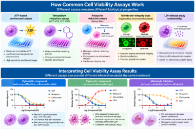
A cytostatic compound may reduce ATP or metabolic reduction signals because cells stop proliferating or alter their metabolism while membrane integrity remains largely intact. Conversely, an acute membrane-disrupting treatment may produce substantial LDH release before the full reduction in ATP or cell number is detected.
For this reason, viability data should be reported according to the assay-specific endpoint rather than treated automatically as a direct count of living cells unless the method has been validated against a cell-counting approach for the experimental model.
Cell Viability Assay Comparison by Readout
| Assay Type | Primary Readout | Major Advantage | Important Consideration |
|---|---|---|---|
| ATP-based | Intracellular ATP | High sensitivity and broad dynamic range | ATP can change with metabolic state; typically an endpoint assay |
| MTT | Tetrazolium reduction | Familiar and economical | Insoluble formazan requires solubilization |
| MTS/XTT/WST | Tetrazolium reduction | Soluble products simplify workflow | Chemical and optical interference can affect results |
| Resazurin | Cellular reducing activity | Sensitive; fluorescence or absorbance detection | Signal depends on metabolic activity and incubation conditions |
| Live/dead dyes | Membrane integrity | Direct visualization and spatial information | Imaging and analysis can increase complexity |
| LDH release | Loss of membrane integrity | Useful for detecting cytolytic injury | Background LDH and assay interference require appropriate controls |
No single assay in this table is universally superior. The most appropriate method depends on the biological endpoint, sample type, instrumentation, throughput requirements, and experimental design.

ATP-Based Luminescent Assays
ATP assays typically use a luciferase-based reaction to generate luminescence related to the amount of ATP present in the sample. They are commonly selected for microplate screening because the signal can be highly sensitive, has a broad usable range under optimized conditions, and is generally suitable for relatively low cell numbers.
The main interpretive consideration is that ATP is a cellular energy marker rather than a direct measurement of membrane integrity. Mitochondrial inhibitors, nutrient limitation, and treatments that alter cellular energy metabolism can change ATP levels before or independently of cell death.
Many commonly used ATP assay formats lyse cells during measurement, making the assay an endpoint measurement that cannot be repeatedly followed in the same well.
ATP assays can be particularly useful when the objective is sensitive relative quantitation of treatment-associated changes across many samples. White, opaque plates are commonly used to improve luminescence collection and reduce optical cross-talk between wells.
Tetrazolium Reduction Assays
MTT, XTT, MTS, WST-1, and related assays use tetrazolium salts that are reduced by metabolically active cells to form colored formazan products. The resulting signal is measured by absorbance.
These assays are widely used because they are relatively economical and compatible with standard microplate readers. However, their practical differences are important.
MTT Assays
MTT produces an insoluble formazan product that typically requires a solubilization step before absorbance measurement. This additional step increases assay time and can introduce well-to-well variation if the crystals are not completely dissolved.
MTT remains useful for established workflows in which assay conditions have been optimized for the particular cell model and treatment.
MTS, XTT, and WST-Based Assays
MTS, XTT, and many WST-based reagents generate water-soluble formazan products, simplifying the workflow by eliminating the crystal-solubilization step required for MTT.
Assay performance still depends on cell type, cell density, incubation time, reagent chemistry, and the reducing capacity of the cells.
Colored test compounds, phenol red-containing media, and particles that scatter light may interfere with absorbance measurements. Some compounds can also interact directly with assay chemistry.
When chemical interference is plausible, include cell-free wells containing the test compound and assay reagent.
Resazurin Reduction Assays
Resazurin-based assays use a blue, weakly fluorescent redox indicator that is reduced to pink, fluorescent resorufin by metabolically active cells. Signal can be measured by absorbance or fluorescence, with fluorescence generally providing greater sensitivity.
Because resazurin reduction reflects cellular metabolic activity, a change in signal does not necessarily demonstrate irreversible loss of cell viability. Treatments that alter cellular metabolism may change the readout even when membrane integrity remains intact.
Extended incubation or very high cell densities can also move the reaction outside its useful linear range. Cell number, incubation time, and assay conditions should therefore be optimized for each cell model.
Resazurin assays may be useful when cells need to remain available after measurement, but reagent exposure and downstream compatibility should be evaluated experimentally. If cells will subsequently be used for protein extraction, microscopy, or another endpoint, a small compatibility study can help determine whether the preceding assay influences the subsequent measurement.
Membrane-Integrity Dyes and Live/Dead Imaging
Trypan blue and related dye-exclusion methods distinguish cells with intact membranes from cells with compromised membranes. These methods are straightforward for suspended cells or adherent cells that can be detached and counted.
Automated cell counters can reduce operator-dependent variability, although cell clumping, debris, and inappropriate threshold settings can still affect results.
Fluorescent live/dead assays commonly combine a cell-permeant fluorescent probe with a membrane-impermeant dye that enters cells after loss of membrane integrity.
These methods provide spatial information that plate-based metabolic assays cannot. They can be particularly useful for imaging-based phenotypic studies, co-cultures, organoids, and experiments in which cell distribution or morphology contributes to interpretation.
The principal trade-off is increased analysis complexity. Fluorescence overlap, uneven staining, background fluorescence, and inconsistent image segmentation can affect quantitation. For adherent cells, however, imaging preserves morphological and spatial information that may be lost after cell detachment.
LDH Release Cytotoxicity Assays
Lactate dehydrogenase release assays detect LDH released into the culture medium following loss of plasma membrane integrity.
LDH is therefore best interpreted as a cytotoxicity measurement rather than as a universal measure of cell viability.
Spontaneous LDH release can increase in stressed cultures, overgrown wells, or mechanically disturbed samples. Serum and other media components may contribute background, while some test compounds can interfere with LDH activity or the coupled detection chemistry.
Appropriate controls are essential. These generally include background or low controls, vehicle-treated controls, and a maximum-release control for determining the total releasable LDH under the assay conditions.
Match the Assay to the Experimental Question
The biological question should determine the assay.
If the objective is to determine whether a compound reduces the number of metabolically active cells in a 96-well screen, ATP, resazurin, or soluble tetrazolium assays may be appropriate.
If the question is whether a treatment causes acute membrane damage, LDH release or a membrane-impermeant fluorescent dye is more directly aligned with that endpoint.
If treatment may arrest proliferation without immediately causing cell death, pair a metabolic readout with cell counting, imaging, DNA-content measurement, or another independent measurement of cell number or proliferation.
For apoptosis and cell viability studies, a viability result alone may not provide sufficient mechanistic information. Early apoptotic cells can retain membrane integrity and measurable metabolic activity. Combining a viability assay with caspase activity, Annexin V labeling, DNA fragmentation, or appropriate protein markers can help distinguish growth inhibition, apoptosis, and later-stage loss of membrane integrity.
Controls and Validation Determine Assay Quality
A reliable cell viability experiment begins with appropriate controls.
Untreated cells and vehicle controls establish baseline behavior, but additional controls may be required depending on the assay format.
Include blank wells containing medium and assay reagent, particularly for absorbance and fluorescence assays. When compounds may interact directly with the assay chemistry or optical readout, include cell-free wells containing the test compound and assay reagent.
Positive controls should reflect the biological endpoint being measured. A membrane-disrupting treatment is appropriate for validating LDH release or dead-cell dye assays, whereas a treatment known to alter metabolic activity may be useful for evaluating metabolic readouts.
Plate layout also matters. Evaporation and temperature gradients can produce edge effects, particularly in multiwell plates. Distributing controls appropriately across the plate can help identify positional variation.
Before comparing treatments, establish a cell-seeding density that remains within the assay’s linear response range at the planned endpoint. Confirm that the vehicle concentration does not independently affect the selected readout.
For dose-response experiments, inspect the raw data as well as normalized percentages. An apparently acceptable dose-response curve can conceal signal saturation, high background, or compound-specific interference.
Common Sources of Cell Viability Assay Error
Several experimental factors can produce misleading viability results:
- Cell density outside the linear range of the assay
- Unequal cell seeding between wells
- Edge effects caused by evaporation or temperature differences
- Test-compound color or fluorescence
- Direct chemical interaction between the compound and assay reagent
- Excessive assay-reagent incubation
- Phenol red or other media-related optical interference
- High spontaneous LDH release
- Cell clumping during dye-exclusion counting
- Changes in metabolism that occur independently of cell death
- Inappropriate normalization or insufficient controls
These effects are one reason assay validation should be performed with the actual cell model, plate format, treatment conditions, and detection instrument used in the experiment.
Practical Selection for Research Workflows
Instrumentation, throughput, sample availability, and downstream requirements should all be considered when selecting a cell viability assay.
Luminescence supports sensitive, high-throughput endpoint measurements.
Absorbance-based assays are practical for laboratories using standard microplate readers and established colorimetric workflows.
Fluorescence assays can provide greater sensitivity and, when combined with imaging, additional spatial and morphological information.
Cell counting and imaging can help distinguish changes in cell number from changes in metabolic state.
LDH assays provide a complementary measure of membrane damage and can be particularly useful when cytolytic injury is the biological endpoint of interest.
The most informative viability experiment is often based on more than one biological property. A scalable primary assay can be followed by confirmation of selected conditions using a method based on a different cellular endpoint.
For example, a metabolic viability assay may be complemented by cell counting or membrane-integrity analysis. This orthogonal approach can help prevent metabolic suppression, cell-cycle arrest, and membrane damage from being interpreted as equivalent biological outcomes.
Supporting Cell Biology Research
Cepham Life Sciences provides research products supporting cell biology and cell-based research workflows, including cell viability and cytotoxicity products, apoptosis research products, cell culture reagents, biological buffers and reagents, laboratory supplies, and related research applications.
For Research Use Only.
References
- Mosmann T. Rapid colorimetric assay for cellular growth and survival: application to proliferation and cytotoxicity assays. Journal of Immunological Methods. 1983;65(1-2):55-63. DOI: 10.1016/0022-1759(83)90303-4)
- O’Brien J, Wilson I, Orton T, Pognan F. Investigation of the Alamar Blue (resazurin) fluorescent dye for the assessment of mammalian cell cytotoxicity. European Journal of Biochemistry. 2000;267(17):5421-5426. DOI: 10.1046/j.1432-1327.2000.01606.x
- Weyermann J, Lochmann D, Zimmer A. A practical note on the use of cytotoxicity assays. International Journal of Pharmaceutics. 2005;288(2):369-376. DOI: 10.1016/j.ijpharm.2004.09.018
- Riss TL, Moravec RA, Niles AL, et al. Cell Viability Assays. Assay Guidance Manual. Bethesda (MD): Eli Lilly & Company and the National Center for Advancing Translational Sciences; 2013, updated 2016.